Reliable imaging support for accurate diagnosis, treatment planning and monitoring across a wide range of medical conditions.
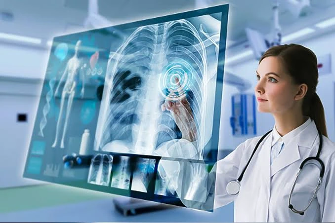

Comprehensive diagnostic imaging services to support accurate evaluation of medical, surgical and emergency conditions.
Specialist-led image interpretation focused on accurate diagnosis, timely reporting and coordinated patient care.
Imaging studies reviewed and interpreted under qualified radiology expertise.
Imaging helps doctors identify disease, injury and structural abnormalities more clearly.
Radiology supports Orthopaedics, Surgery, Urology, Gynaecology, Vascular Care and other departments.
Investigations selected according to the clinical condition and individual diagnostic requirement.
Radiology uses medical imaging to help diagnose, evaluate and monitor diseases and injuries affecting different parts of the body.
At Chiranjeevi Hospitals, radiology supports specialists across multiple departments by providing imaging information needed for accurate diagnosis and treatment planning.
Depending on the clinical condition, imaging may be used to evaluate bones, joints, abdominal organs, kidneys, blood vessels, soft tissues and other internal structures.
The appropriate investigation is selected according to symptoms, examination findings and the referring doctor’s clinical requirement.
Radiologists interpret imaging findings and work with treating specialists to provide clinically relevant diagnostic information.
The goal is to support timely diagnosis, appropriate treatment decisions and continued monitoring where required.
Medical imaging may be recommended when symptoms or examination findings require a clearer view of internal organs, bones, joints, blood vessels or soft tissues.
Doctors may advise imaging for persistent abdominal pain, injuries, swelling, suspected stones, joint problems, vascular conditions or other unexplained symptoms.
Imaging is also commonly used to confirm a diagnosis, assess the severity of a condition, plan treatment or monitor recovery.
The appropriate investigation depends on the clinical question. Not every patient requires the same type of scan, so imaging should be selected according to medical need.
Radiology supports diagnosis across a wide range of medical and surgical conditions.
Imaging helps evaluate fractures, arthritis, injuries and other musculoskeletal abnormalities.
Diagnostic imaging can help assess the liver, gallbladder, pancreas, spleen and other abdominal structures.
Imaging may help identify kidney stones, obstruction and other urinary-system abnormalities.
Doppler imaging can evaluate blood flow within arteries and veins.
Pelvic imaging may assist in evaluating the uterus, ovaries and related structures.
Imaging helps assess selected swellings, masses and soft-tissue abnormalities.
Chest imaging may help evaluate lung, pleural and other thoracic abnormalities.
Imaging can assist doctors in assessing injuries and acute medical conditions.
The imaging investigation is selected according to symptoms, clinical findings and the area requiring assessment.
Commonly used to evaluate bones, joints, chest conditions and selected injuries.
Uses sound waves to examine abdominal organs, pelvic structures and other soft tissues.
Evaluates blood flow within arteries and veins and helps identify selected vascular abnormalities.
Provides detailed cross-sectional images for selected medical, surgical and emergency conditions when required.
Provides detailed soft-tissue imaging and may be advised for selected neurological, musculoskeletal and other conditions.
Ultrasound or other imaging guidance may assist selected diagnostic procedures when clinically appropriate.
Radiology services are selected according to the patient’s clinical condition and the diagnostic information required.
Diagnostic imaging for bones, joints, chest and selected medical conditions.
Non-invasive imaging for abdominal, pelvic, soft-tissue and other selected conditions.
Ultrasound-based assessment of blood flow in arteries and veins.
CT or MRI may be advised when more detailed anatomical evaluation is clinically required.
Imaging assessment to support the diagnosis of injuries and acute medical or surgical conditions.
Repeat imaging may be used to monitor disease progression, healing or response to treatment when required.

Consultant Radiologist
MBBS – Gold Medalist, MD Radiology
Medical imaging plays an important role in modern healthcare by helping doctors see structures and abnormalities that cannot always be assessed through physical examination alone.
Different imaging methods provide different types of information. X-rays are commonly used for bones and chest evaluation, while ultrasound can assess abdominal organs, pelvic structures and blood flow without ionizing radiation.
More detailed cross-sectional imaging may be required for selected conditions depending on the clinical question.
Radiologists interpret these images in combination with the patient’s medical information and provide findings that help treating doctors determine the next step.
Patients from Kukatpally, KPHB, JNTU, Nizampet, Miyapur, Moosapet and nearby Hyderabad areas can access Radiology services at Chiranjeevi Hospitals.
Ultrasound uses high-frequency sound waves to produce images of structures inside the body without using ionizing radiation.
It is commonly used to evaluate abdominal organs, kidneys, urinary structures, pelvic organs and selected soft tissues.
Colour Doppler ultrasound adds information about the direction and pattern of blood flow within arteries and veins.
Doppler evaluation can therefore support the assessment of vascular conditions such as reduced arterial circulation, venous problems and selected blood-flow abnormalities.
The type of ultrasound or Doppler study recommended depends on the symptoms and clinical condition being investigated.
Radiologist interpretation helps combine the imaging findings with the clinical question so that the treating doctor receives meaningful diagnostic information.
A radiologist is a medical specialist who interprets diagnostic imaging and provides findings that help other doctors diagnose and manage medical conditions.
Imaging can help evaluate fractures, organ abnormalities, stones, vascular problems, soft-tissue conditions and many other medical concerns.
X-rays use ionizing radiation and are commonly used for bones and chest imaging, while ultrasound uses sound waves to visualize soft tissues and internal organs.
Colour Doppler is an ultrasound technique used to evaluate blood flow through arteries and veins.
Ultrasound is generally a non-invasive and well-tolerated diagnostic examination.
Preparation depends on the type of examination. Some abdominal or pelvic scans may require fasting or a full bladder.
No. The appropriate imaging test depends on symptoms, examination findings and the clinical information required.
You can access Radiology services and consult Dr. Pavani, Consultant Radiologist, at Chiranjeevi Hospitals, KPHB Phase 1, Kukatpally, Hyderabad.
Get appropriate diagnostic imaging and specialist radiology interpretation to support accurate evaluation and treatment planning.
Diagnostic imaging evaluated under qualified radiology expertise.
Imaging services supporting medical, surgical and emergency departments.
Investigations selected according to individual clinical requirements.
Clear imaging information to assist diagnosis and treatment planning.
Our expert team is ready to provide personalized care for you and your family. Book your appointment today and take the first step towards better health.

©2026 Chiranjeevi Hospitals