Modern medicine relies entirely on accurate information. When you experience sudden pain, a traumatic injury, or unexplained internal symptoms, a doctor cannot simply guess what is wrong. Before any surgical incision is made, any cast is set, or any oncology treatment is prescribed, your medical team must be able to “see” exactly what is happening deep inside your body. This is where the profound science of medical imaging becomes the most critical step in your healthcare journey.
Radiology is the technological backbone of a modern hospital. It provides the crucial roadmap that guides surgeons, oncologists, neurologists, and emergency physicians. Without precise, high-resolution imaging, accurate diagnosis is impossible, and treatment becomes a dangerous game of trial and error.
To ensure our patients receive the most accurate diagnoses possible, Chiranjeevi Hospitals offers a state-of-the-art Radiology Department providing comprehensive, high-resolution diagnostic imaging and interventional services in Hyderabad. Whether you require an emergency CT scan for trauma at our KPHB branch or an advanced whole-body MRI at our newly opened Kondapur branch, our department is fully equipped with cutting-edge technology and expert radiologists to provide rapid, precise, and safe diagnostic care.
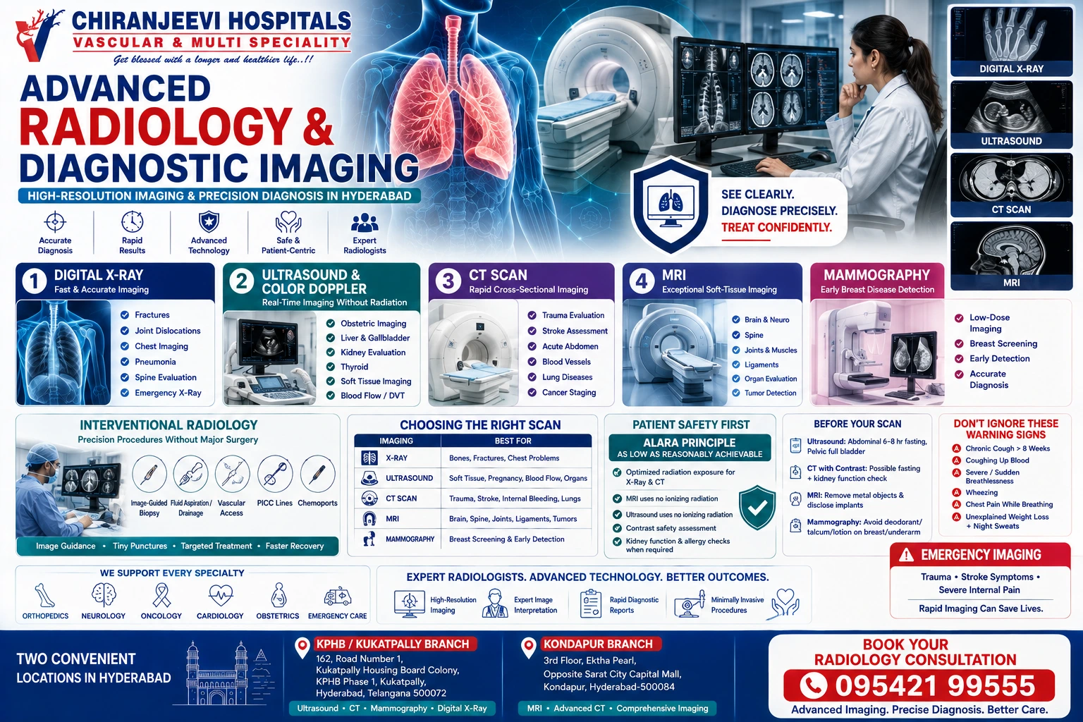
The Radiology Department at Chiranjeevi Hospitals provides a complete spectrum of diagnostic and image-guided clinical services. Our core services include high-resolution Magnetic Resonance Imaging (MRI), multi-slice Computed Tomography (CT Scans), advanced 3D/4D Ultrasound and Color Doppler, Digital X-Rays, and routine Mammography for breast cancer screening. Furthermore, we offer specialized Interventional Radiology services for image-guided biopsies, fluid aspirations, and minimally invasive vascular procedures. Utilizing strict radiation safety protocols, our department delivers exceptional, patient-centric imaging services across both our KPHB and Kondapur branches.
Direct Answer: The Radiology Department is a specialized medical and technological wing dedicated exclusively to acquiring and interpreting medical images to diagnose and treat diseases. We unify advanced imaging technology with expert sub-specialized radiologists to provide crucial diagnostic reports that guide all other medical departments, from emergency trauma care to complex oncology.
A common misconception is that radiology is just a room where “pictures” are taken. In reality, a Radiology Department is a highly complex clinical environment. The radiologist is a specialized medical doctor trained to interpret intricate shades of gray, anatomical variations, and subtle tissue differences to identify diseases at their earliest, most curable stages.
At Chiranjeevi Hospitals, our radiology services are structured around two critical clinical pillars:
Diagnostic Radiology: This focuses on using non-invasive imaging technologies to look inside the body and identify the cause of a patient’s symptoms. Diagnostic services are used to screen for diseases (like breast cancer), monitor how well a body is responding to a treatment (like shrinking tumors after chemotherapy), and diagnose acute emergencies (like a stroke or a ruptured appendix).
Interventional Radiology (IR): This is the minimally invasive surgical wing of the department. Interventional radiologists use real-time imaging (like live X-ray fluoroscopy or ultrasound) to guide tiny catheters, wires, and needles deep into the body through blood vessels or small punctures. This allows them to perform highly targeted treatments—such as draining deep abscesses, performing core biopsies, or stopping internal bleeding—without the need for traditional open surgery.
Key Takeaway: The Radiology Department touches almost every patient who walks into the hospital. By housing advanced imaging technology and expert interpreters under one roof, we ensure that your treating physician receives a rapid, highly accurate diagnosis to formulate your treatment plan without dangerous delays.
Direct Answer: Our Radiology Department utilizes a comprehensive suite of imaging modalities, each serving a unique medical purpose. The most frequently utilized services include Digital X-Ray for bone evaluation, Ultrasound for soft tissue and obstetrics, CT Scans for trauma and complex structures, and MRI for incredibly detailed neurological and joint imaging.
Not all imaging machines do the same thing. Each technology uses a different physical mechanism—sound waves, radiation, or magnetic fields—to visualize specific types of tissue.
Digital Radiography (X-Ray): The most fundamental and widely used imaging tool. X-rays use a very small, safe dose of ionizing radiation to create images of dense structures. They are the immediate gold standard for diagnosing bone fractures, joint dislocations, and evaluating the chest for pneumonia or fluid in the lungs. Digital X-rays offer significantly lower radiation doses and instant image availability compared to older film techniques.
Ultrasonography (USG) & Color Doppler: Ultrasound uses high-frequency sound waves (echoes)—not radiation—to create real-time images of the body’s internal organs. It is entirely safe for pregnant women and is widely used for obstetrics (fetal monitoring), evaluating the liver, gallbladder, and kidneys, and examining the thyroid. Color Doppler is a specialized ultrasound feature that allows us to visualize and measure the speed of blood flow through veins and arteries, crucial for detecting deep vein thrombosis (DVT).
Computed Tomography (CT Scan): A CT scanner takes a series of X-ray images from different angles around your body and uses computer processing to create highly detailed, cross-sectional slices (like slices of a loaf of bread) of the bones, blood vessels, and soft tissues. It is incredibly fast, making it the primary modality for emergency trauma, acute strokes, appendicitis, and detailed cancer staging.
Magnetic Resonance Imaging (MRI): MRI uses a massive, powerful magnetic field and radio waves—no ionizing radiation—to generate exceptionally detailed 3D images of soft tissues that do not show up well on X-rays. It is the absolute gold standard for imaging the brain (detecting tumors or multiple sclerosis), the spinal cord (herniated discs), and complex joints (torn ligaments or meniscus injuries in the knee).
Mammography: A specialized, low-dose X-ray system designed exclusively for examining the breast tissue. It is the primary diagnostic service used for the early detection and diagnosis of breast diseases in women, capable of revealing tiny microcalcifications and masses years before they can be felt during a physical exam.
Direct Answer: Interventional Radiology (IR) utilizes real-time imaging to perform minimally invasive procedures. Using ultrasound or CT guidance, our radiologists can perform precise tissue biopsies, drain deep internal fluid collections, and place vital central venous lines, often utilizing only a needle puncture rather than a surgical incision.
Interventional Radiology has revolutionized modern medicine. By substituting tiny cameras and image guidance for large surgical incisions, IR services drastically reduce a patient’s pain, lower the risk of infection, and cut recovery times from weeks to mere hours.
Key Interventional Services provided by our department include:
Image-Guided Biopsies: When a suspicious mass is detected on a scan (e.g., in the liver, lung, or breast), a piece of that tissue must be extracted to test for cancer. Instead of cutting the patient open, an interventional radiologist uses live ultrasound or CT scans to safely guide a tiny needle directly into the mass to extract the cells precisely, avoiding surrounding blood vessels and organs.
Fluid Drainage (Aspiration): Infections or diseases can cause dangerous fluid to pool in the abdomen (ascites) or around the lungs (pleural effusion), causing severe pain and breathlessness. Under ultrasound guidance, our radiologists can safely insert a tiny catheter to drain this fluid and provide immediate relief.
Vascular Access: For patients requiring long-term chemotherapy, dialysis, or intravenous antibiotics, IR specialists use ultrasound to safely thread special long-term IV lines (like PICC lines or Chemoports) into the large central veins, sparing the patient from painful, repeated needle sticks in their arms.
Direct Answer: Patient safety is our highest priority. We strictly adhere to the ALARA principle, As Low As Reasonably Achievable ensuring radiation doses for X-rays and CT scans are kept to the absolute minimum. When contrast dyes are necessary to highlight blood vessels, we utilize advanced, kidney-safe agents and monitor patients closely for any rare allergic reactions.
Many patients express anxiety about radiation exposure and the use of intravenous “dye.” Understanding the safety protocols of our Radiology Department can alleviate these fears.
Radiation Safety: While X-rays and CT scans do use ionizing radiation, modern digital equipment is incredibly efficient, requiring only a fraction of the radiation used a decade ago. The medical benefit of accurately diagnosing a life-threatening condition like an internal bleed or a tumor exponentially outweighs the negligible risk of a single diagnostic scan. Ultrasounds and MRIs use zero ionizing radiation.
What is Contrast Dye? For many CT and MRI scans, the radiologist may request a contrast agent. This is a special liquid (usually iodine-based for CT, and gadolinium-based for MRI) injected into your vein. It briefly travels through your bloodstream, causing blood vessels, tumors, and inflamed tissues to “light up” brightly on the scan, making them easily distinguishable from normal tissue.
Contrast Safety: Before administering contrast, our clinical team will always check your kidney function (via a Serum Creatinine blood test) to ensure your kidneys can safely filter and flush the dye out of your body. We will also ask about your allergy history to prevent any adverse reactions.
| Common Myth | Medical Fact |
| Myth: An MRI scan exposes you to massive amounts of dangerous radiation. | Fact: An MRI uses a strong magnetic field and radio waves to create images. It uses absolutely zero ionizing radiation and is completely safe in terms of radiation exposure. |
| Myth: Ultrasounds are only used to look at babies during pregnancy. | Fact: While crucial for obstetrics, ultrasounds are widely used to diagnose gallstones, kidney blockages, deep vein thrombosis (blood clots), and thyroid nodules in both men and women. |
| Myth: A CT scan and an MRI are basically the exact same thing. | Fact: They use different technology and excel at different things. CT is superior for imaging dense bone, acute bleeding, and the lungs. MRI is far superior for imaging soft tissues like the brain, spinal cord, and ligaments. |
| Myth: If you are claustrophobic, you cannot have an MRI. | Fact: While MRI machines are enclosed, our department offers open communication, soothing techniques, and, if necessary, mild sedation (prescribed by a physician) to help claustrophobic patients complete their scans comfortably. |
To ensure your scan produces the highest quality diagnostic images, patient cooperation and preparation are essential. Different modalities require different preparations:
For Ultrasounds: If you are having an abdominal ultrasound, you will generally be asked to fast (eat or drink nothing) for 6 to 8 hours before the scan to reduce intestinal gas, which blocks sound waves. For a pelvic ultrasound, you will be required to drink plenty of water and hold your urine so your bladder is completely full, which acts as a “window” to see the uterus and ovaries clearly.
For CT Scans with Contrast: You will likely be asked to fast for 4 hours prior to the test. Ensure you bring your latest Serum Creatinine blood test report, as the technologist must verify your kidney function before administering the IV contrast dye.
For MRIs: The MRI room contains a massive, incredibly powerful magnet. It is constantly on. You must remove all metallic objects—including jewelry, watches, hairpins, hearing aids, and undergarments with metal underwires. You must strictly inform the technologist if you have a cardiac pacemaker, aneurysm clips, or any metal implants in your body, as the magnet can violently pull or heat these objects.
For Mammograms: Do not wear deodorant, talcum powder, or lotion under your arms or on your breasts on the day of the exam. These substances can appear as white spots on the X-ray, mimicking calcifications and causing false alarms.
The clinical circumstances surrounding diagnostic imaging vary immensely. One patient may require a routine outpatient ultrasound to monitor a pregnancy, while another may present in the emergency room following a severe car accident, requiring a rapid, full-body trauma CT scan to locate internal bleeding. Assessment and image acquisition must be swift, highly individualized, and expertly interpreted.
At Chiranjeevi Hospitals, patients gain access to a fully equipped Radiology Department that prioritizes rapid imaging, high-resolution diagnostic clarity, and strict patient safety protocols.
Guided by our expert team of Consultant Radiologists and skilled radiologic technologists, the department offers a seamless blend of routine screening, advanced disease staging, and emergency interventional care. From routine chest X-rays to complex, multi-phase CT angiograms, our services are designed to provide your treating physicians with the exact clinical roadmap they need to protect your health and save your life.
(Note: A routine diagnostic appointment is not appropriate for acute emergencies. A person experiencing severe trauma, symptoms of a stroke (facial drooping, weakness), or sudden, excruciating internal pain should seek immediate emergency medical attention at our 24/7 casualty department, where emergency imaging will be prioritized.)
To ensure specialized diagnostic services and advanced imaging are highly accessible, Chiranjeevi Hospitals operates fully equipped Radiology Departments across two strategic locations in Hyderabad. Both branches offer comprehensive diagnostic screening and expert radiological interpretations.
| Location | Suitable For | Nearby Catchment Areas |
| KPHB / Kukatpally Branch | Patients in northwest/west Hyderabad seeking expert diagnostic ultrasound, CT scans, mammography, and routine X-ray services. | KPHB, Kukatpally, JNTU, Nizampet, Miyapur, Moosapet |
| Kondapur Branch | Patients in western Hyderabad and the IT corridor looking for advanced MRI services, complex CT imaging, and comprehensive diagnostic evaluations. | Kondapur, Hitech City, Madhapur, Gachibowli, Hafeezpet |
Chiranjeevi Hospitals — KPHB / Kukatpally Branch
Location: 162, Road Number 1, Kukatpally Housing Board Colony, KPHB Phase 1, Kukatpally, Hyderabad, Telangana 500072
Chiranjeevi Hospitals — Kondapur Branch
Location: 3rd Floor, Ektha Pearl, Opposite Sarat City Capital Mall, Kondapur, Hyderabad-500084
Contact & Appointments
Appointment Booking: [Visit Chiranjeevi Hospitals Contact & Booking Page]
Department Profile: [View Our Radiology Department Services]
Phone / WhatsApp Consultations: (Insert official hospital contact number here)
The Foundation of Diagnosis: Accurate medical treatment begins with accurate imaging. The Radiology Department provides the essential visual data required by every other medical specialty.
Right Tool for the Job: CT scans excel at imaging bone, lungs, and trauma, while MRIs are the gold standard for soft tissues like the brain, spinal cord, and ligaments.
Radiation Safety: We adhere to strict ALARA principles, utilizing advanced digital equipment to minimize X-ray and CT radiation doses, while MRI and Ultrasound use zero radiation.
Interventional Precision: Our Interventional Radiology services allow us to perform critical biopsies and fluid drainages through a needle puncture, avoiding traditional surgical incisions.
Two Accessible Locations: Comprehensive diagnostic imaging, advanced CT/MRI services, and routine X-rays are available at both our established KPHB branch and our newly opened Kondapur branch.
Facing an unexplained illness or a sudden injury is highly stressful, and the uncertainty of not knowing what is happening inside your body only amplifies that anxiety. Massive advancements in diagnostic imaging technology mean that modern medicine no longer relies on guesswork. With the ability to visualize the human body in high-resolution, 3D cross-sections, radiologists can detect diseases earlier and map out surgical interventions with pinpoint accuracy. The key to effective treatment is partnering with a fully equipped medical department that utilizes state-of-the-art technology alongside expert clinical interpretation. By trusting the advanced Radiology Services at Chiranjeevi Hospitals, you gain access to the clarity, precision, and diagnostic certainty necessary to chart your path to recovery.
1. What radiology services are available at the Kondapur branch?
The new Chiranjeevi Hospitals Kondapur branch offers a comprehensive suite of diagnostic imaging services. This includes advanced MRI scans, multi-slice CT scanning, high-definition 3D/4D ultrasound and Color Doppler, and digital X-ray services for both emergency and outpatient evaluations.
2. Can I eat before an ultrasound scan?
It depends on the type of ultrasound. For an abdominal ultrasound (checking the liver, gallbladder, etc.), you must fast for 6 to 8 hours beforehand to reduce bowel gas. For a pelvic ultrasound (checking the uterus or bladder), you do not need to fast, but you must drink plenty of water to ensure a full bladder.
3. Do I need a doctor’s prescription to get an MRI or CT scan?
Yes. High-level diagnostic imaging such as CT scans and MRIs require a valid clinical prescription (referral) from a licensed medical practitioner. This ensures that the correct scan and protocols (such as whether or not to use contrast dye) are selected based on your specific medical symptoms.
4. Where is the Radiology Department located in Hyderabad?
Comprehensive Diagnostic Imaging and Radiology services are highly accessible through our two major Chiranjeevi Hospitals facilities: our established primary center in KPHB Phase 1 (Kukatpally) and our new, modern branch located in Kondapur.
5. Which Chiranjeevi Hospitals branch is convenient for patients from Hitech City seeking an MRI?
For patients residing or working in Hitech City, Madhapur, and Gachibowli, the Radiology Department at the Chiranjeevi Hospitals Kondapur branch offers the most convenient access to advanced MRI services and complex diagnostic imaging.
6. Which branch is convenient for patients from Kukatpally requiring a routine X-ray or Ultrasound?
Patients located in Kukatpally, JNTU, Nizampet, and Miyapur will find the digital X-ray, Mammography, and Ultrasound services at the Chiranjeevi Hospitals KPHB branch extremely convenient for their routine diagnostic evaluations.
7. How long does an MRI take?
Unlike a CT scan, which takes only a few seconds, an MRI is a longer process. Depending on the body part being scanned, an MRI typically takes between 20 to 45 minutes. You will be required to lie completely still during this time to prevent the images from blurring.
8. Are X-rays safe for pregnant women?
Generally, medical professionals avoid exposing the abdominal and pelvic areas of pregnant women to X-rays or CT scans unless it is an absolute life-threatening emergency. If imaging is required during pregnancy, Ultrasound or MRI (without contrast) are the preferred, safe modalities. Always inform the technologist if there is any chance you might be pregnant.
Radiological Society of North America (RSNA) Patient Information Guidelines
American College of Radiology (ACR) Appropriateness Criteria
Indian Radiological and Imaging Association (IRIA) Clinical Standards
The information provided in this article is for educational and informational purposes only and describes the services offered by the hospital. It should not be construed as medical advice. Diagnostic imaging must be prescribed by a licensed medical practitioner based on a clinical evaluation. Always consult your doctor before undergoing any radiological procedure, and strictly follow all pre-scan preparation instructions provided by the imaging department.
Our expert team is ready to provide personalized care for you and your family. Book your appointment today and take the first step towards better health.

©2026 Chiranjeevi Hospitals